- Details
- Category: Light microscopy
- Hits: 2293
The Leica STED-CW is the complementary device to the confocal microscope providing nanoscopic research level. The device allows to study living cells with the dot pattern of 512 x 512 dots, with the up to 18 frames/sec speed and with the resolution up to 80 nm; to study living cells a number of fluorescen proteins could be used, such as Citrine, YFP, etc.; to study fixed material with the resolution up to 80 nm conventional fluorescent dyes such as Alexa 488, FITC and Oregon Green could be used; to use singe molecule detection technique for the reduction of the excitation spot in the lateral plane up to 80 nm. For more information visit www.leica-microsystems.com.

- Details
- Category: Light microscopy
- Hits: 2324
Leica WLL is the complementary system to the confocal microscope allowing fine tuning of fluorochrome excitation wavelengths resulting in the minimum cross-excitation and specimen damage. The system provides spectral specimen scanning with the excitation wavelength acquiring; phototoxicity reduce; fluorescence attenuation imaging (FLIM) with the excitation within 470-670 nm; precise spectral discrimination of fluorochromes with nearby emission spectra; etc. For more information visit www.leica-microsystems.com.

- Details
- Category: Light microscopy
- Hits: 2142
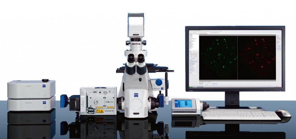
The Cell Observer Spinning Disk (Nipkow Disk) confocal microscope is designed to study quickly proceeding processes inside the living cell. In contrast to the scanning laser confocal microscopy, spinning disk confocal microscopy allows to reach high speed scanning with decent contrast and minimized phototoxicity. The instrument provides all types of applications requiring sensitivity and speed in the trade-off the resolution, such as high-speed imaging of the dynamic processes in cells, time-lapse imaging with low phototoxicity, etc. For more information visit microscopy.zeiss.com.

- Details
- Category: Light microscopy
- Hits: 2186
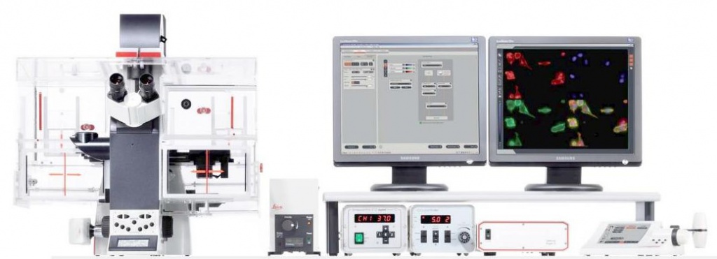
The Leica DMI6000 is an inverted fluorescence microscope equipped with microhandling system providing micromanipulations and structured illumination system OptiGrid providing ultra-high-contrast. The microscope allows to perform dual excitation ratiometric investigations with two-excitation maximum fluorescent dyes such as FURA2, BCECF; transmitted light sample analyses with different types of contrasting, such as bright field, dark field, phase-contrast, polarization, integrated modulation contrast, differential interference contrast; epifluorescence sample analysis; 3D-fluorescence imaging; etc. For specifications visit www.leica-microsystems.com.

- Details
- Category: Light microscopy
- Hits: 1947
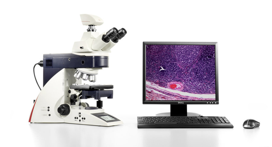
The Leica DMI4000 is a research microscope allowing to perform a wide range of science-research investigations with different contrasting methods. It can be equipped with customers accessories based on application needs. For more information visit www.leica-microsystems.com.


