• About Us
    • How to Apply for Research Studies
    • Application for measurement for external users
    • Regulations
      • Consent to the processing of personal data
  • Research Park
    • Resource Centres
    • Equipment
    • Methods
    • Service list
  • News
  • Work of Research Park
    • Reviews
    • Publication
    • Statistics
  • Promotional materials
    • Our partners
    • Presentations
    • Booklet Research Park
  • Contacts

ATOMIC-FORCE MICROSCOPY

Description of the types of bacteria


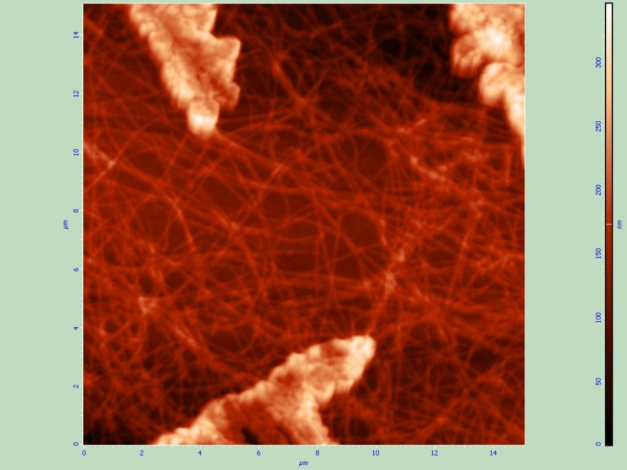
High-resolution chromosome analysis and collagen fibers


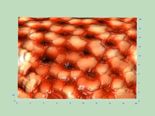
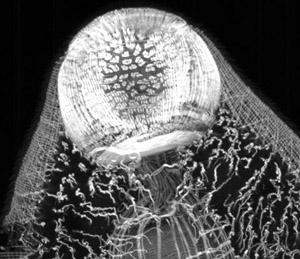
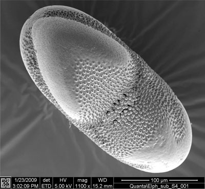
The morphology of surface section of the diatoms


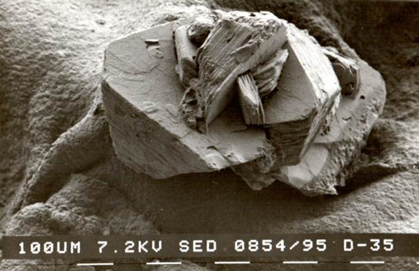
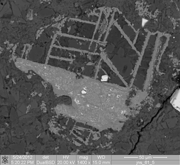
Morphology of diamond crystals


LASER SCANNING CONFOCAL MICROSCOPY

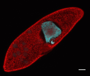
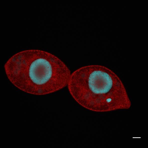
Studying bacteria in ciliates as a possible source of human pathogenic bacteria
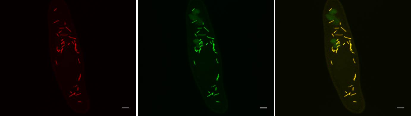
Ciliates


Visualization muscle actin in marit. 


SCANNING ELECTRON MICROSCOPY AND ELECTRON-PROBE MICROANALYSIS

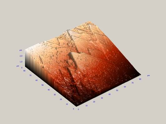
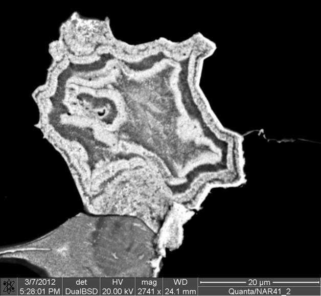
The morphology of of mineral aggregates.


The morphology of shells of diatoms.


Studying malicious ticks-fitoparasites.


Phase contrast, which depends on the density (average atomic number) of the irradiated material.


Energy dispersive X-ray spectra, scored at different points of the studied material.


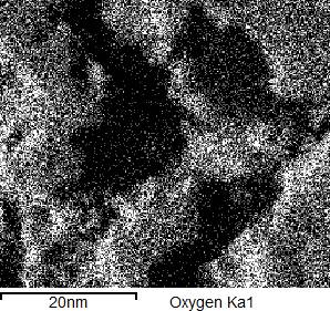
Maps of the distribution of chemical elements, obtained by scanning the sample surface.


Select your language

  • Russian
  • English (UK)
  • About Centre MM
  • Equipment RC MM
  • Methods RC MM
  • Staff RC MM
  • Contacts RC MM
  • Website RC MM
© 2025 Research Park, St Petersburg University
Support: esrc-support@spbu.ru