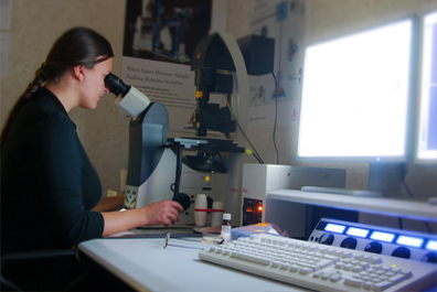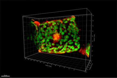
Laser scanning microscopy based on Leica TCS SP5 microscope equipped with resonant scanner. The device can accommodate specimens labeled with all fluorophores including ultra violet. High-speed imaging supplies the data for a wide range of integrated analytical techniques.

High-resolution light and fluorescence microscopy using laser scanning microscope Leica TCS SP5, automated Leica fluorescence microscopes, equipped with high resolution monochromatic and color digital cameras, cube filters for wide range of fluorochromes and lenses for phase- and DIC-contrast. Complex also includes stereomicroscopes for microsurgery and preparation.

Electron microscopy technique based on comprises transmission electron microscope Tesla BS-500, ultra-sectioning devices and glass-knife maker.

Molecular and cell biology probe preparation based on complex of devices, such as centrifuges, electrophoresis set, gel detector, PCR thermocyclers, hybridizers, microinjector Nanoject II, spectrophotometer, PCR and laminar boxes and cryotome-cryostat.

Image processing for the analysis of microscopy results based on specialized software Bitplane Imaris, Autoquant X3, Huygens Professional for performing bioimaging: 3D-reconstruction, deconvolution and statistical analysis.


